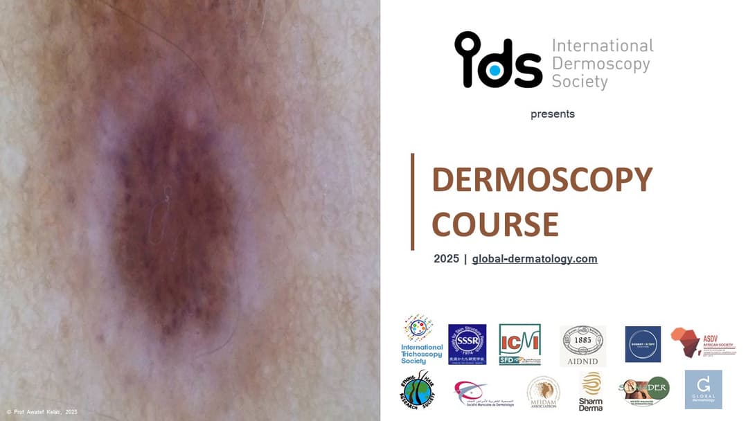
Topographic Dermoscopy
4h50
1 modules
17 lectures
Accreditation

Topographic Dermoscopy

Chair:Prof. Awatef Kelati, MD, PhD
Topographic Dermoscopy
4h50
1 modules
17 lectures
Accreditation
Topographic Dermoscopy

Dermoscopy in Facial lesions: Melanocytic facial lesions
Elvira Moscarella MD, PhD

Dermoscopy in chest and back lesions: Skin tumors
Cristian Navarrete-Dechent MD, PhD

Dermoscopy of conjunctival lesions
Elisa Cinotti MD, PhD

Dermoscopy in lesions of the limbs
Bengu Nisa Akay MD, PhD
Dermoscopy of the mucosae: Genital mucosae
Zoe Apalla MD, PhD

Dermoscopy in Facial lesions: Inflammatory dermatoses of the face
Enzo Errichetti MD, MSc, DVD
Dermoscopy in lesions of the palms and soles
Raimond Karls MD
Dermoscopy of the ear
Awatef Kelati MD, PhD
Dermoscopy in Facial lesions: Non melanocytic pigmented lesions, Fair skin
Ashfaq Marghoob MD, PhD

Dermoscopy in Facial lesions: The nose
Sebastian Podlipnik MD

Dermoscopy in haïr and scalp disorders: Tumors of the scalp
Susana Puig MD, PhD

How do not to miss nail unit cancer
Luc Thomas MD, PhD
Dermoscopy in haïr and scalp disorders: Diffuse alopecia
Antonella Tosti MD
Scabies mite is bright green under U.V dermatoscopy
Pawel Pietkiewicz MD
Dermoscopy in chest and back lesions: Inflammatory and infectious skin diseases
Gabriel Salerni MD, MSc, PhD

Dermoscopy in Facial lesions: Non melanocytic pigmented lesions, Dark skin
Balachandra Ankad MD, MBBS

Dermoscopy in Facial lesions: Non melanocytic pigmented lesions, Dark skin. Part 2
Vinay K MD
Learning objectives
Topographic dermoscopy is an advanced approach in dermatology that adapts dermoscopic examination techniques to specific anatomical locations, recognizing that skin morphology, appendage distribution, and environmental exposure significantly influence dermoscopic patterns. This method enhances diagnostic precision by integrating regional context into the interpretation of dermoscopic features.
On the face, dermoscopy reveals distinct characteristics due to the high density of pilosebaceous units, thin epidermis, and chronic sun exposure. Common findings include a pseudonetwork caused by interruption of pigmentation around follicles, annular-granular structures, and yellowish globules. Facial lentigines, actinic keratoses, and early lentigo maligna each present region-specific clues that differ from their counterparts on the trunk or limbs.
The auricular region (ear) poses particular challenges due to its concave and convex contours and sebaceous-rich but hairless skin. Dermoscopy in this area helps differentiate between benign lesions such as seborrheic keratoses and high-risk entities like lentigo maligna or basal cell carcinoma, which often show linear irregular vessels, shiny white structures, or asymmetric pigmented networks.
On the chest and back, where the skin is thicker and often exposed to intermittent sun exposure, dermoscopic patterns may include structureless pigmentation, regression structures, or atypical pigment networks. Special attention is needed for large congenital or acquired nevi, as well as for early melanoma that may not yet present classical dermoscopic criteria.
Lesions on the limbs are influenced by friction, mechanical trauma, and variable vascularization. Dermoscopy in these regions often shows polymorphous vessels, globules, or clods with pigmentation patterns that may mimic malignancy, especially in areas of chronic irritation.
The palms and soles (acral regions) require specific dermoscopic criteria due to their unique ridge and furrow anatomy. The parallel ridge pattern is a hallmark of melanoma, while the parallel furrow, lattice-like, and fibrillar patterns are more common in benign melanocytic nevi. Understanding the alignment of pigmentation with skin furrows or ridges is key to accurate diagnosis.
Scalp and hair disorders are assessed using trichoscopy, a specialized form of dermoscopy that identifies follicular openings, peripilar signs, hair shaft anomalies, and perifollicular changes. Common trichoscopic features include yellow dots in alopecia areata, miniaturized hairs in androgenetic alopecia, and comma hairs or corkscrew hairs in tinea capitis.
Nail disorders benefit from onychoscopic evaluation, especially when assessing longitudinal melanonychia, hemorrhages, nail matrix pigmentation, or dystrophic changes. Dermoscopy distinguishes between benign pigmentation (e.g., ethnic melanonychia or nail matrix nevi) and alarming features such as Hutchinson’s sign or irregular pigment bands indicative of subungual melanoma.
Mucosal dermoscopy provides additional diagnostic support for pigmented and vascular lesions of the lips, oral cavity, and genitalia. Although challenging due to moisture and mobility, mucoscopy can reveal blue-white veils, multiple colors, or irregular vessels in suspicious mucosal melanomas or vascular tumors.
An emerging adjunct to topographic dermoscopy is ultraviolet dermoscopy. Under UV light, certain organisms and structures fluoresce, aiding in diagnosis. For instance, scabies mites appear as bright green dots under UV dermoscopy, allowing rapid confirmation of infestation even in atypical presentations.
Overall, topographic dermoscopy enhances dermatological diagnostics by tailoring interpretation to regional anatomy, improving sensitivity and specificity, and guiding targeted management strategies.
Might interest you
PsychodermatologyDermoscopyNeglected Tropical Disease
Cutaneous Medicine: Multidisciplinary Approaches in Dermatology
Chair: Prof. Fahafahantsoa Rapelanoro Rabenja,
This course explores the intersection of dermatology with other medical specialties, emphasizing a collaborative approach to diagnosing and managing complex skin disorders. It covers a wide range of topics, including dermatopathology, rheumatology, oncology, and infectious diseases, highlighting how systemic conditions manifest cutaneously. With contributions from experts in various fields, the text provides comprehensive insights into multidisciplinary care, advanced diagnostic techniques, and innovative treatments. Ideal for dermatologists, internists, and specialists, it bridges gaps between disciplines to improve patient outcomes in cutaneous medicine.

Pigmentation
Pigmentation
Chair: Dr Seemal Desai, MD, FAAD
Hyperpigmentation is excess skin color from melanin. Understand melanin synthesis mechanisms and main causes.
Neglected Tropical Disease
Neglected Tropical Skin Diseases
Chair: Dr. Prajwal Pudasaini, MD
Neglected tropical skin diseases affect poor populations in tropical areas. They include leprosy, mycetoma, and cutaneous leishmaniasis, causing disability and stigma. They receive little attention and resources, leading to poor diagnosis and treatment. Increased awareness and improved healthcare access are needed to help affected communities.

TCM Chinese Medicine
Acne Treatment in China
Chair: Prof. Haiping Zhang, PhD
Acne treatment in China combines traditional methods with modern practices.
Pigmentation
Cyspera Medical Education
Chair: Global Dermatology,
Cyspera® is a topical pigment-correcting treatment formulated with cysteamine, a naturally occurring compound that reduces the appearance of persistent hyperpigmentation, including melasma, post-inflammatory hyperpigmentation, and lentigines. It is known for being non-hydroquinone, suitable for long-term use, and effective on all skin types.
Topographic Dermoscopy
Chair: Prof. Awatef Kelati, MD, PhD
Topographic dermoscopy refers to the region-specific application of dermoscopic examination, emphasizing the unique morphological patterns found across different anatomical sites. On facial skin, the dermoscopic assessment requires recognizing patterns influenced by the high density of pilosebaceous units and sun-induced changes, often presenting pseudonetworks and annular-granular structures. The ear, with its thin skin and sebaceous gland concentration, reveals specific vascular and follicular clues important in distinguishing benign from malignant lesions.
On the chest and back, where the skin is thicker and sun exposure varies, dermoscopy must account for irregular pigment distribution and architectural disorder, especially in large nevi or early melanomas. Limb lesions may show distinctive features due to mechanical friction, hair density, and vascular variations, demanding precise interpretation to identify atypical nevi or skin cancers.
Palmar and plantar dermoscopy highlights the parallel ridge pattern critical for melanoma diagnosis, contrasting with benign acral patterns like the parallel furrow or lattice-like structures. Scalp and hair disorders benefit from trichoscopy, where dermoscopic evaluation reveals specific signs such as yellow dots, broken hairs, or black dots, aiding in the diagnosis of alopecia areata, androgenetic alopecia, or tinea capitis.
In nail disorders, onychoscopy enables visualization of melanonychia, hemorrhages, and nail matrix changes, crucial for distinguishing subungual melanoma from benign causes like trauma or fungal infection. Mucosal dermoscopy, though technically challenging, provides diagnostic clues in pigmented lesions of the lips, genitalia, or oral mucosa, requiring adaptation to moist, non-keratinized surfaces.
Finally, ultraviolet dermoscopy reveals a unique application: scabies mites fluorescing bright green under UV light, enhancing detection when traditional visualization fails. Topographic dermoscopy thus demands both anatomical knowledge and technical adaptation to maximize diagnostic accuracy across diverse body sites.
Acne
ACNE
Chair: Dr. Jerry Tan, MD
The ACNE | Education Series, led by Dr. Jerry Tan, is a comprehensive global medical education initiative designed to enhance the understanding and skills of dermatologists and healthcare practitioners regarding acne. Participants will gain insights from leading international experts on the latest advancements in acne research, innovative treatment options, and patient-centered care approaches. The event features interactive discussions, live Q&A sessions, and evidence-based strategies, all at no cost. The esteemed faculty includes specialists from the USA, Italy, France, the UK, Singapore, Greece, Australia, Canada, and Germany. This is a valuable opportunity to improve clinical competencies and stay updated on current acne management practices.
Attendees will acquire up-to-date knowledge on acne pathophysiology, new therapeutic options, and patient-oriented management strategies to optimize clinical outcomes in acne treatment. The session will also provide practical insights through expert-led discussions and evidence-based approaches.
Psychodermatology
Psychodermatology
Chair: Prof Mohammad Jafferany, MD
This specialized course explores the vital intersection between dermatology and mental health, equipping clinicians with the knowledge and tools to manage psychodermatological conditions effectively. Through a blend of theoretical knowledge and practical application, participants will learn to diagnose and treat dermatological delusional disorders, identify psychiatric comorbidities in skin disease patients, and implement mental health strategies for chronic dermatoses. The curriculum also examines psychological factors in cosmetic dermatology and provides cutting-edge screening techniques for body dysmorphic disorder, including modern digital manifestations like Zoom dysmorphia. Adopting a patient-centered approach, the course emphasizes multidisciplinary management of conditions where psychological and dermatological factors interact. Participants will gain expertise in recognizing psychiatric components of skin diseases, addressing the emotional
burden of chronic conditions, and applying ethical principles in cosmetic practice. The training combines expert instruction with case-based learning to bridge theory and clinical practice. Designed for dermatologists, psychiatrists, psychologists, and primary care providers, this program enhances clinicians' ability to deliver holistic care that addresses both the visible and invisible aspects of skin disorders. Upon completion, practitioners will be better prepared to manage complex psychodermatological cases while improving patient outcomes through integrated mind-skin healthcare.
Neglected Tropical Disease
Tropical Dermatology and Neglected Tropical Dermatoses
Chair: Prof. Fahafahantsoa Rapelanoro Rabenja,
Dermatological diseases, particularly neglected tropical diseases (NTDs) with skin manifestations like deep mycosis (chromoblastomycosis, sporotrichosis, mycetoma), scabies, leprosy, lymphatic filariasis, and cutaneous leishmaniasis, pose major challenges for healthcare systems in resource-limited regions of Africa, Asia, and Latin America. These conditions severely affect vulnerable populations, suffering from frequent underdiagnosis and inadequate treatment that exacerbates suffering. Diseases such as atopic dermatitis are also under consideration for inclusion as skin NTDs through collaborative efforts involving ISAD, ASDV, and WHO. Furthermore, albinism, highly prevalent in sub-Saharan Africa, presents significant social challenges including stigmatization and occult beliefs. Despite these complex difficulties, the field is undergoing a historic transformation driven by science and technology, particularly artificial intelligence (AI), which offers tangible tools for improving diagnosis, treatment, and prevention. The participation of global experts facilitates vital knowledge exchange, exploration of innovative solutions, and helps address critical shortages of human and material resources in remote areas.
Learning Objective:
Understand the complex challenges posed by dermatological diseases, especially skin NTDs and conditions like albinism, in resource-limited settings, and recognize the critical role of global collaboration, technological innovation (particularly AI), and expert knowledge exchange in developing solutions to improve diagnosis, treatment, prevention, and resource allocation.
Dermoscopy
Dermoscopy
Chair: Prof Awatef Kelati, MD, PhD
This comprehensive dermoscopy course provides dermatologists and healthcare professionals with essential skills in skin lesion evaluation, covering fundamental principles through advanced diagnostic applications across five key areas: global dermoscopy practices, pigmented lesion analysis (including differentiation of benign and malignant patterns), specialized techniques for skin of color, skin cancer detection (melanoma and non-melanoma), and general dermatological conditions (inflammatory, infectious, and hair/nail disorders). Participants will develop proficiency in recognizing diagnostic patterns, adapting techniques for diverse skin types, and applying dermoscopic algorithms, ultimately enhancing their clinical accuracy through a combination of theoretical knowledge and practical case-based learning. The course emphasizes real-world application, addressing both common and challenging scenarios in dermatological practice.